- SELF STUDY MODULES
- 1. Intro to TBI
- 2. Communication
- 3. Skills for independence
- 4. Cognitive changes
- 5. Behaviour changes
- 6. Sexuality
- 7. Case management (BIR)
- 8. No longer available
- 9. Mobility & motor control
- 10. Mental health & TBI:
an introduction - 11. Mental health problems
and TBI: diagnosis
& management - 12. Working with Families
after Traumatic Injury:
An Introduction - 13. Goal setting
1.3 Describe the basic anatomy of the skull and brain
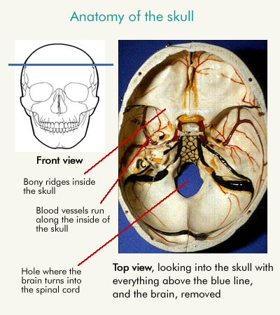
i) The skull
The skull is a hard, bony box protecting the brain. It fits neatly around the brain, with the brain sitting inside, floating in cerebrospinal fluid.
There are a number of bony ridges that are on the inside of the skull that fit into gaps in the brain. These ridges can cause damage to brain tissue in the event of a TBI. This damage can commonly include lacerations or contusions around the frontal/ temporal lobes and contra-coup damage in the region of the occipital lobe.
Blood vessels line the inside of the skull, and there is a hole (the foramen magnum) at the base of the skull where the brain becomes the spinal cord.
ii) The brain: Introduction
The brain is made up of two different types of tissue:
- Grey matter: which is groups of nerve cells that form layers of soft grey tissue
- White matter: which is long fibres that connect different groups of cells and have the consistency of al dente spaghetti or jelly
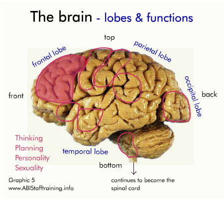
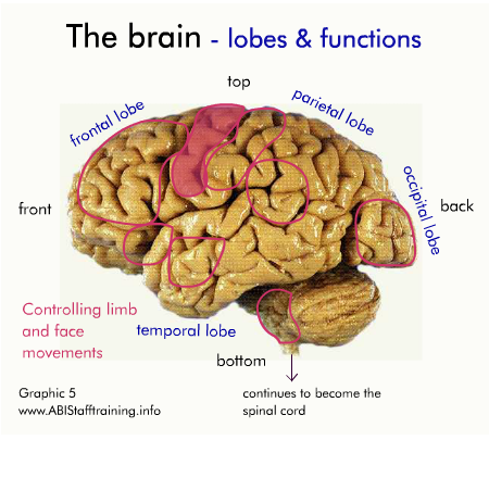
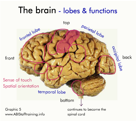
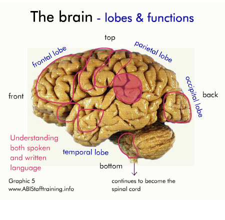
The brain has a left and right hemisphere. Each hemisphere controls the opposite side of body. The left side also usually controls understanding and production of speech. Each hemisphere is divided into 4 lobes - frontal, temporal, parietal, occipital. Different lobes, and different areas within each lobe, control different functions.
At the base of the brain, just above where it turns into the spinal cord is an area called the brain stem. The brain stem controls our vital functions such as breathing. If the brain stem is damaged it can be extremely life threatening.
SLIDES: To pause: Hover mouse over slide. To continue: move mouse off slide.
To go to a specific slide: Click on slide numbers below.

Many of the things we do depend on several functions, which means several of these areas must work together. For example, in order to follow the direction "put your socks on" you need to use the following areas:
- the area for understanding language, in order to understand the direction
- the area for spatial orientation, to be able to find your sock
- the area for controlling limb movements, in order to put your sock on
- and the area for planning in order to be able to plan these steps and execute them in the correct order
To allow the different areas of the brain to work together, the brain has networks of nerve fibers that connect the areas together. There are also nerve fibers that connect the brain to the rest of the body, allowing it to control the activities of the person. If either the area that controls a function, or the nerve fibers that connects that area to the rest of the brain or to the body are damaged, a person will have difficulty performing that function.
People often focus on exercise or physiotherapy for an arm or leg that doesn't work, and think the problems is with the limb, when it may be the part of the brain that controls that limb, or the nerve fibers that connect the brain to the limb that have been damaged.
iii) The brain: Lobes and hemispheres
The brain is the body's control centre. It may only weigh about 1.5kg but it is made up of billions of cells. It controls everything we do from basic bodily functions such as breathing, heart beat and blood pressure, to our movements, speech, senses and our personality.
Cerebral Hemispheres
The brain is divided into two cerebral hemispheres – the left hemisphere and the right hemisphere. These hemispheres are largely symmetrical in size and structure. The two hemispheres work seamlessly together, processing sensory information from the environment and initiating motor responses.
Generally speaking, the movement on the right side of our body (the actions of our arms and legs) is controlled by the left side of the brain (left hemisphere) and vice versa, movement on the left side of the body is controlled by the right side of the brain (right hemisphere).
Each hemisphere also has specialised functions. The left hemisphere tends to play a significant role in speech and language. The right hemisphere plays a special role in the perception and integration of nonverbal information including facial recognition.
Lobes
The cortex of each hemisphere comprises four ‘lobes’ that handle specific areas of function. The animation immediately below shows the left and right hemispheres and the four lobes (red, green yellow and light yellow) within each hemisphere. (The blue area is the cerebellum.)
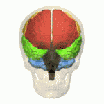
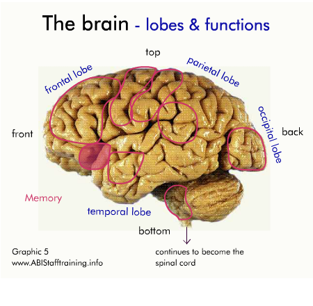
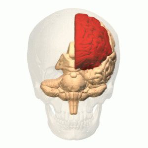
Frontal lobes
The frontal lobes have different subdivisions. The back division of the frontal lobes are part of the brain’s systems for controlling our movement. The front division of the frontal lobes play an important role in higher-level thinking skills (such as planning, organising, reasoning, decision-making and judgement.), as well as aspects of behaviour, emotions and personality. They rely on complex connections and information exchanges with other areas of the brain to perform these ‘executive functions’. The animation immediately below shows the left frontal lobe.
Parietal lobes
The parietal lobes are divided into two zones. The front half of the parietal lobes are principally concerned with receiving data from the bodies’ senses such as touch, pressure, temperature, and pain and then communicating this information to the frontal lobes. The rear section of the parietal lobes deals with spatial awareness, such as our ability to find our way around, and to reach for objects. Interestingly, this region also plays an important role in our ability to do arithmetic.
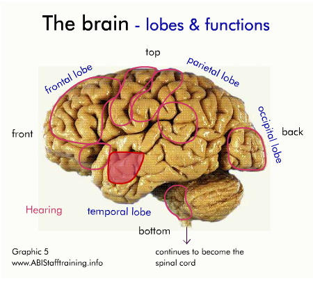
Temporal lobes
The temporal lobes also have a role in language, particularly in the ability to hear and understand. The temporal lobes are also concerned with memory, the emotions, the ability to enjoy music and to recognise and identify things we see, such as faces or objects.
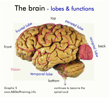
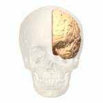
Occipital lobes
The occipital lobes are primarily concerned with vision but also with our ability to recognise what we see in terms of identifying colours, locating objects in the environment and seeing objects accurately.
The animation immediately below shows the left occipital lobe.
Cerebellum and Brain Stem
Below the cerebral hemispheres are the cerebellum and the brain stem, which connect with the spinal cord.
Cerebellum
The cerebellum ‘carries out’ orders from the cerebral hemispheres and keeps a number of vital but routine functions kicking over, such as maintaining balance and ensuring our muscles move in a smooth, coordinated way.
Brain stem
The brain stem controls many vital functions including breathing, blood pressure, blood circulation, swallowing, appetite, body temperature and digestion, as well as the need for water, staying awake and sleeping, among other things. It is also the main route for nerve fibres running between the cerebral hemispheres and the spinal cord. Even small injuries to the brain stem can be life threatening.
![]()